- >>
- Healthy Adult Studies
- HCP Young Adult
- News
- Article
NY Times articles provide an "inside-the-scanner" look at HCP

Although many start their careers as scientists, it’s rare that a science reporter actually gets to participate in a scientific project. Last summer, New York Times reporter Jim Gorman got that chance, and wrote about it in two articles published today in the New York Times.
The Human Connectome Project hosted Gorman and his videographer at Washington University in St. Louis to get first hand experience with what it feels like to be a participant in the project and shoot a video about the experience.
In the article “The Brain, in Exquisite Detail”, the reporter profiles the project through conversations with HCP investigator Deanna Barch, director of the team that guides participants through the battery of in-scanner and out-of-scanner tests used for HCP. In the video, Dr. Barch explains:
What we’re doing in this project is pretty different in a couple [of] ways. We have really state-of-the-art techniques and equipment that are going to let us do this in a much finer-grained way than has ever been done before.
We’re going to be studying approximately 1,200 individuals across a wide range of things like education levels, and income levels, people from different racial and ethnic backgrounds, so we can have a much better sense of the kind of “true normal” range of brain connections …
Some of it is just basic science, trying to understand how the brain works and how the brain contributes to how we behave, but a lot of it has clinical application.
In “A Search for Self in a Brain Scan”, Gorman offers a more personal, and philosophical angle on what it is like to be scanned and, ultimately, to be able to look at images of your own brain.
Gorman was treated to several hours of MRI scanning, physical, behavioral, and cognitive tests, just as if he were one of the 1,200 participants being scanned for the HCP. Between scans, he talked with HCP investigators and research assistants about what he was experiencing, what can be learned from the data we’re collecting, and the significant effort required to process the data and make it available to the public.
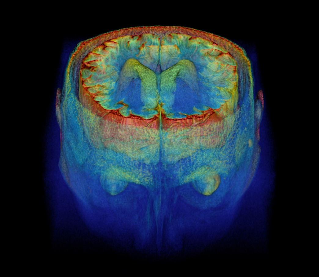
A few examples of detailed brain visualizations afforded from the high resolution of the HCP MRI scans are featured, along with Jim Gorman’s journey through the scanner, in the article’s video: