- >>
- Healthy Adult Studies
- HCP Young Adult
- News
- Article
Multi-scale Connectomics: HCP in <em>Science</em> technologies feature
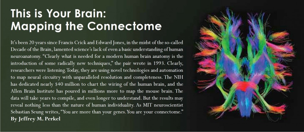
This month, Science magazine spotlights efforts to map the neural connectomes at the macroscopic, mesoscopic, and microscopic levels in a Life Science Technologies special feature by Jeffrey M. Perkel. The article is part of series meant to provide readers with “information on leading edge lab products and useful career advice and trends”.
The article outlines the technology being developed to map the whole brain (macroscopic) human connectome by HCP consortia based at WU-UMinn and at MGH-UCLA. Kamil Ugurbil, Co-PI of our own WU-UMinn HCP, is quoted in the article:
Kamil Ugurbil, director of the Center for Magnetic Resonance Research at the University of Minnesota and co-PI of the WashU/Minnesota consortium, says his team has seen “significant technological gains” with their new scanner—resolution has been increased two- to three-fold and some 30 subjects have already been scanned, each over a two-day period.
Efforts at the Allen Institute for Brain Science and Cold Spring Harbor Laboratory (Partha Mitra) to map neuronal pathways from regions in the mouse brain through axonal projections (the “mesoscopic” connectome) and the technology necessary to do this are detailed. The article ends by highlighting specialized, automated electron microscopy developed by Jeff Lichtman at Harvard and crowd-sourcing efforts by Moritz Helmstaeder at the Max Planck Institute of Neurobiology to trace neuron-to-neuron connections that make up the human retinal connectome at the microscopic scale.
The amazing technologies necessary for deciphering brain connections shows just how true the observation Francis Crick and Edward Jones made in 1993 was:
Clearly what is needed for a modern human brain anatomy is the introduction of some radically new techniques.